Vascular Doppler
Vascular Doppler is an imaging test that involves the use of high-frequency soundwaves to assess and evaluate a blockage or obstruction in the arteries and veins, especially those supplying oxygenated blood to the limbs. Vascular Doppler can help to detect a wide gamut of diseases and problems concerning blood vessels including blood clots. This helps to determine the risks of having a stroke and take necessary measures if the patient falls in the risk category.The procedure is performed as a part of a blood flow study and does not require any major preparation.
Uses of Carotid Doppler
Vascular Doppler can help to detect, evaluate and assess the following problems:
- Obstruction in the arteries which may be a result of plaque build-up
- Blood clot formation in the blood vessels
- Hampered or poor circulation of blood which may be a result of serious damage to the blood vessels
- Venous occlusion
- Spastic arterial disease
- Blockage or clotting in the prosthetic artery after artificial bypass grafting.
Indications of Vascular Doppler
Doctors usually recommend Vascular Doppler to patients with any of the following problems:
- Deep vein thrombosis (DVT)– The condition is marked by the formation of blood clots in the deep veins which are usually present in the limbs which may be the result of an injury or side-effect of previous surgery.
- Superficial Thrombophlebitis – The condition is marked by severe inflammation in the veins which results due to the formation of blood clots right beneath the skin surface.It commonly occurs in the legs.
- Arteriosclerosis – the condition is marked by narrowing or stiffening of the arteries which are responsible for carrying oxygenated blood to the limbs. It is likely to affect elderlies but can be found in younger people as well.
- Thromboangiitis Obliterans – Commonly known as Buerger disease, it is a quite rare disease marked by the recurring inflammation in blood vessels present in the hands and feet.
- Vascular Tumours – Benign or malignant growths and lesions in the blood vessels.
Working
The procedure involves the use of an ultrasound scanner which consists of a computer console, display screen and a transducer, which is a small portable device which helps to generate high-frequency soundwaves. These waves bounce back on striking the tissues and vessels, producing echoes. The transducer not only produces the sound waves but also records the waves that are echoed back. The same are converted to images that are generated on the display screen.
Preparing for the procedure
As such there are no mandatory guidelines that need to be followed before a Vascular Doppler. The patient might, however, be asked to refrain from smoking for several hours prior to the test. Also if you are on any medication, ask your doctor whether you need to temporarily discontinue taking these.
During the procedure
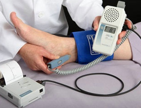
The patient is made to lie down on his/her back on the examination table. A special gel will be applied on the concerned area to reduce the amount of air between the skin and the transducer. In order to monitor and compare the blood pressure in various parts of a body like the thighs, ankles, calf, and arms, pressure cuffs may be used. The transducer will be gently placed against the skinand moved to and forth. The patient might feel slight pressure. The images, thus generated, are recorded and used to evaluate the arteries and veins.
Benefits of Vascular Doppler
- The procedure is non-invasive which means no needles and no pain
- The procedure is very simple and does not require any prior preparation
- The use of soundwaves makes the procedure extremely safe for even children and pregnant women.
- It helps to generate precise and clear images, which is not always possible with normal x-rays.
Risks
The procedure is completely risk-free as it does not involve any incision or medication.
Limitations of Vascular Doppler
- Although Vascular Doppler is highly effective for evaluating superficial vessels, deep vessels might not be clearly visible and require more detailed tests like CT (computerised tomography) scan and MRI (magnetic resonance imaging).
- It is slightly difficult to evaluate smaller blood vessels as compared to the larger ones.
- Calcifications which result from chronic atherosclerosis obstruct the ultrasound beams thereby preventing these from reaching the blood vessels and tissues.